“Keeping PACE with Your Health”
It comes as no surprise that most men often ignore warning signs and symptoms when it comes to their health.

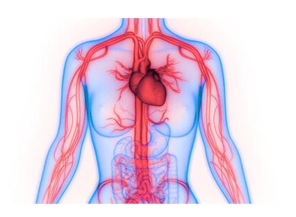
Angiography was first used as a radiological treatment in the 1920s. Using a hollow needle, contrast agent, and live radioscopy, doctors were able to scan vessels. At the time, the only technical choices were to implant the needle into a vessel and inject the contrast agent straight through the same needle. As a result, not all target vessels could be successfully scanned.
With the passage of time, this notion expanded, expanding the technological possibilities. Vessel imaging grew more selective, even for vessels that were less accessible, such as those in the belly. In addition, a growing number of approaches for endovascular therapy of pathologic diseases have been established. This principle has now given rise to a wide range of approaches. The premise of vascular interventions will be discussed in this article, using a variety of commonly utilized vascular treatments as an example.
Vascular intervention may be necessary for a variety of causes. This will be examined in further depth in the next sections. Other specialties, such as surgery, suggest patients for intervention, and they notify their patients about the treatment. Outpatient consultations are available at several hospitals' radiology departments. Renal function, coagulation status, and allergies are all examined in order to prepare for the surgery. Because the contrast agent used during the operation is damaging to the kidneys, adequate renal function is required. Additional efforts to safeguard the kidneys are necessary if renal function is impaired (prehydration). It is critical to maintain a good coagulation state in order to avoid bleeding. Anticoagulants are commonly used by patients, and they should be stopped well before the treatment. Although hospital protocols may vary, an INR of less than 2 is commonly tolerated for vascular treatments. Finally, allergies must be considered since some individuals may experience a severe response to the contrast agent or local anesthetic. Anti-inflammatory and antihistaminic drugs must be given prophylactically if this is known.
The vasculature can be examined in a variety of methods when vascular disease is suspected. Echo duplex is the least intrusive and least expensive type of ultrasound, and it's usually employed when a particular issue regarding a small portion of the vasculature has to be answered. However, there are a few disadvantages: Assessment is subjective, time-consuming, and highly dependent on the physical features of the patient. Due to overhanging intestinal loops and extensive adipose content, assessing abdominal veins, in particular, might be difficult. CT or MR angiography is utilized to offer extra information as necessary.
In the angio room, vascular procedures are conducted. It includes a transportable table for the patient, an X-ray machine, and a screen that displays the radioscopy pictures. The target region may be imaged in the chosen direction using this arrangement. Short movies are created instead of X-rays, and all treatments may be followed 'live'. Contrast compounds can enhance imaging of particular tissues (blood arteries, urinary system, bile ducts, and so on).
A blood vessel is pierced with a hollow needle holding a guide wire in this technique. Palpation can be used to guide this, but in the radiology department, ultrasound is frequently employed. The needle is then removed from the vessel, leaving the guide wire in place. The sheath, a thick working tube, is next put over the guide wire. A valve at the end of the sheath prevents blood from leaking through the sheath. The common femoral artery is the most common blood vessel utilized for access. Because to its shallow location and enormous size, access is rather simple. The brachial artery is also commonly employed. The popliteal artery, also known as the crural artery, is rarely utilized. Other arteries, such as the superficial femoral artery, are less suited because to their deep location and the lack of underlying bone to press against when the needle is inserted.
The common femoral vein or the jugular vein are commonly utilized for venous access.
Once the sheath is in place, it gives stable access to the circulatory system, allowing for the passage of various types of guide wires and catheters. These guide wires and catheters may be moved to the desired location using live radioscopy. Guide wires and catheters come in a wide range of shapes and sizes. The length, tip curvature, flexibility, and substance of guide wires can all vary. For blood vessel travel, flexible hydrophilic guide wires are commonly utilized. They are less distressing, but lack steadiness, which is a disadvantage.
In the case of rigid guide wires, the reverse is true. When difficult tissue must be pierced to install a sheath, stiff guide wires give required rigidity. Flexible guide wires are less suited in this situation since there is less "push" available. Catheters come in a variety of sizes and forms. The tip, which may come in a variety of forms, is the most noticeable distinction. The type of catheter used is determined by the blood artery that has to be catheterized. The abdominal arteries split from the aorta at a severe angle, necessitating a catheter with a greater bend than the leg arteries, which are generally straight.
A (flexible hydrophilic) guide wire is always used in front of the catheter to navigate through the blood arteries. Because guide wires are less stressful to the vessels, this is critical. There is a substantially increased risk of problems such as dissection or even perforation of an artery if a catheter is put first. When the target location is reached, the wire is removed from the catheter, and other materials, such as contrast agent for imaging, medicine, or embolization material, can be delivered through the catheter.
The sheath must be removed from the patient after the operation. Because the sheath leaves a huge hole in the vessel, this is not an easy surgery. The hole can be closed by using extended finger compression followed by a pressure bandage. Vascular closure devices have also been developed in recent years.
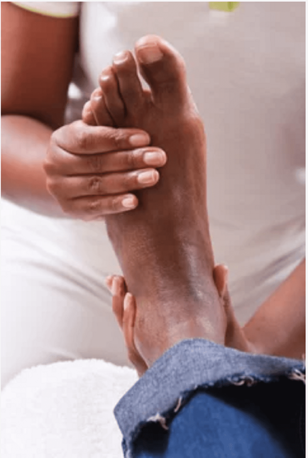
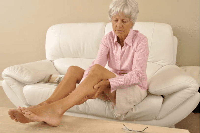
When a closure device is utilized, the concept is that it reduces the danger of additional bleeding, is less uncomfortable for patients, and allows them to move around more readily. As an added bonus, no prolonged pressing is necessary, allowing the angio chamber to be ready for the next patient sooner. The majority of patients who are candidates for vascular intervention have stenosing/occlusive vascular disease, which results in ischemic symptoms. This usually happens in the lower extremities. It is caused by atherosclerosis in the vast majority of instances. Age, smoking, diabetes mellitus, and hypertension are all risk factors for the development of atherosclerosis. Thromboembolic processes, vasculitides, external compression, and other factors can all contribute to stenosing/occlusive vascular disease.
We are just a call or click away. To learn more, book an appointment online or over the phone with PeachState Advanced Cardiac & Endovascular. We have several locations in Georgia: Newnan, Atlanta, & Griffin.